Minimally Invasive Lobectomy
A Modern Approach to Lung Surgery
A Minimally Invasive Lobectomy is an advanced surgical procedure to remove one lobe of the lung through small incisions, without the need for a large, open chest operation. This technique is regularly used to treat early-stage lung cancer but may also be indicated for certain infections, benign lung tumours, or other abnormalities. The minimally invasive method offers significant benefits compared to traditional surgery, including reduced pain, faster recovery, and a shorter hospital stay.
At Mr Pick’s Heart & Lung Practice, we are committed to delivering the least invasive approach to lung surgery possible. Our use of cutting-edge techniques allows us to minimise trauma, protect surrounding tissue, and enhance post-operative comfort and recovery.
Understanding the Lungs and Lung Surgery
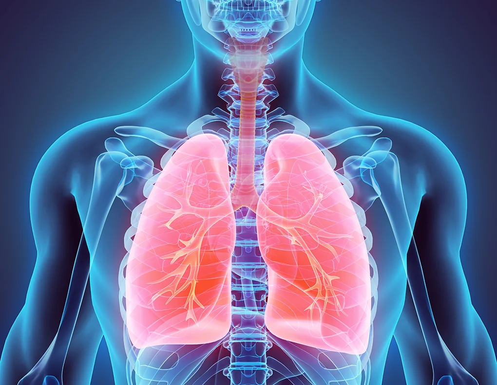
The lungs are housed in the chest cavity and are covered by a thin lining called the pleura, which also lines the inner surface of the rib cage. As we breathe, these linings slide over each other smoothly. The right lung has three lobes, while the left has two. Depending on the condition and extent of disease, surgery may involve removing a part of a lobe (wedge resection or segmentectomy), a full lobe (lobectomy), or even an entire lung (pneumonectomy).
Our Approach: Minimising Invasiveness and Pain
Traditional vs Minimally Invasive Access
Traditional lung surgery involves a thoracotomy—a large 30cm incision in the chest wall, often requiring rib-spreading retractors. At Mr Pick’s practice, we do not use thoracotomies. Instead, we employ a thoracic access port technique, using:
- A single 3-5 cm incision under the arm (along the bra-line in women) to access the lung.
- Thoracoscopy ports—a 1 cm incision that allow insertion of a high-definition camera and surgical tools for a technique known as video-assisted thoracoscopic surgery (VATS). An extrapleural catheter is also introduces to bathe all affected intercostal nerves with anaesthetic solution for 48 hrs post procedure.
How the Procedure Works
Under general anaesthesia, the patient is positioned on their side. One lung is deflated to allow space for surgery, while the other continues breathing via a special tube. Using the VATS approach, small ports are inserted:
- A camera provides a clear internal view of the lung.
- Surgical instruments are guided through the thoracic access port.
- When required, abnormal tissue or the entire lung lobe is removed through the small incision.
After the surgery, drain tubes are inserted through the same small incisions to remove air and fluid from the chest cavity. These are usually removed within a few days.
To further reduce pain, an extra-pleural catheter may be inserted to deliver local anaesthetic directly to the chest nerves for 48 hours, often eliminating the need for additional intravenous or oral painkillers.
Types of Lung Resection Procedures
Wedge Resection
Removal of a small, wedge-shaped portion of lung containing a lesion and a margin of healthy tissue. Often used when lung function is limited. However, the chance of cancer recurrence is higher.
Segmentectomy
A larger portion than a wedge but smaller than a full lobe. May be used for early-stage tumours or to preserve lung function.
Lobectomy (Minimally Invasive)
The complete removal of a lobe of the lung using small incisions and thoracoscopic tools. It is the standard surgical treatment for most early-stage lung cancers and offers the best chance of cure while minimising invasiveness.
Pneumonectomy
Removal of an entire lung. This is only performed when necessary and lung function must be carefully assessed beforehand.
Sleeve Lobectomy
Removal of a lobe along with a segment of bronchus (airway) followed by reattachment of the remaining bronchus to preserve more lung tissue.
Lymphadenectomy
Removal of nearby lymph nodes during surgery to assess and stage possible malignancy.
Why Patients Are Referred for Lobectomy
Most patients come to Mr Pick after imaging, such as a chest x-ray or PET scan, reveals a suspicious lung nodule or mass. Lung cancer risk is higher in individuals with a history of smoking or other risk factors. To ensure the most appropriate treatment:
- A pathologist may be present in theatre to immediately examine biopsy samples.
- Lung function tests (spirometry) are performed before surgery to determine how much lung can be safely removed.
- The goal is always to preserve as much of the healthy lung as possible while ensuring complete removal of the disease.
Risk and Benefits of Minimally Invasive Lobectomy
Benefits:
- Less post-operative pain
- Smaller incisions and less scarring
- Shorter hospital stays
- Faster return to normal activities
- Reduced risk of complications compared to traditional thoracotomy
Potential Risks:
- Bleeding
- Infection
- Air leak from the lung that may take time to heal
- Pneumonia or inflammation of the remaining lung
- Damage to nearby organs or nerves (rare)
- Risks related to general anaesthesia (nausea, allergic reactions, etc.)
A Trusted, Patient-Centred Approach
Mr Pick’s Heart & Lung Practice prioritises minimally invasive techniques and patient comfort. Our approach ensures optimal outcomes with less trauma and quicker recovery. If you’ve been diagnosed with a lung lesion or early-stage lung cancer, we’re here to guide you through your treatment options with clarity and care.